Every orthopedic procedure – whether diagnosis, treatment decision-making, preoperative planning, or surgery – begins with a clear understanding of the patient’s anatomy.
In the case of a joint replacement, before a customized implant can be chosen or designed, manufactured, and validated, there is one essential step: identifying the relevant anatomical structures from medical images. In a nutshell, the surgeon first needs to understand where the bone is, what shape it has, and how it relates to its surrounding anatomy.
This process is known as image segmentation – and, in the context of orthopedics, more specifically, bone segmentation.
For the ADAPT’s project, bone segmentation is a key part of the digital workflow. By transforming medical imaging data into precise anatomical models, segmentation helps to create the foundation for patient-specific implant design, supporting the development of orthopedic implants adapted to each patient’s bone structure. ADAPT’s objectives include the development of AI-based algorithms for bone segmentation, which are then used to support high-precision 3D generative designs for customized implants.
What Is Image Segmentation?
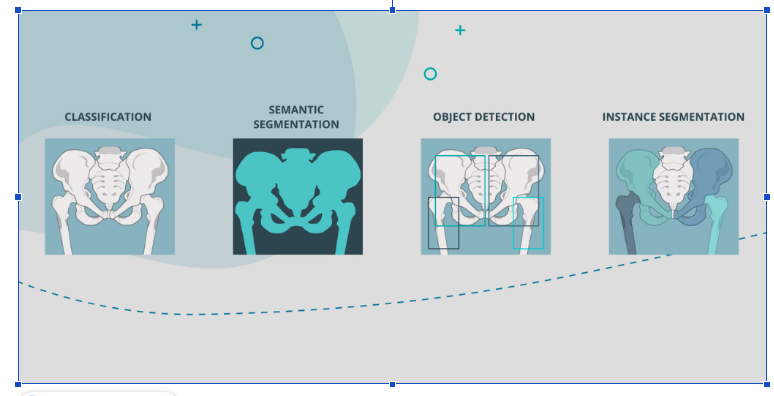
In simple terms, image segmentation is a computer vision technique that divides a digital image into meaningful parts or regions.
Instead of looking at an image as a single block of pixels, segmentation helps identify what each part of the image represents. In medical imaging, this may mean separating bone from soft tissue, identifying different anatomical structures, or outlining a specific region of interest.
In orthopedics, image segmentation can be used to identify and delineate bones and other anatomical structures from imaging exams such as X-rays, CT scans, or MRI scans. This makes it possible to transform raw medical images into structured, clinically relevant information. Medical image segmentation is widely described as a process that identifies and outlines specific structures or regions of interest within diagnostic images, making it particularly useful for pre-operative planning and post-operative assessment.
What Is Bone Segmentation?
Bone segmentation is the application of image segmentation to skeletal anatomy.
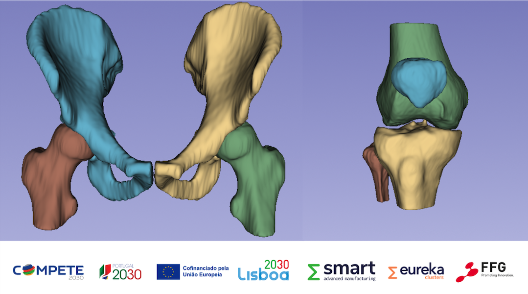
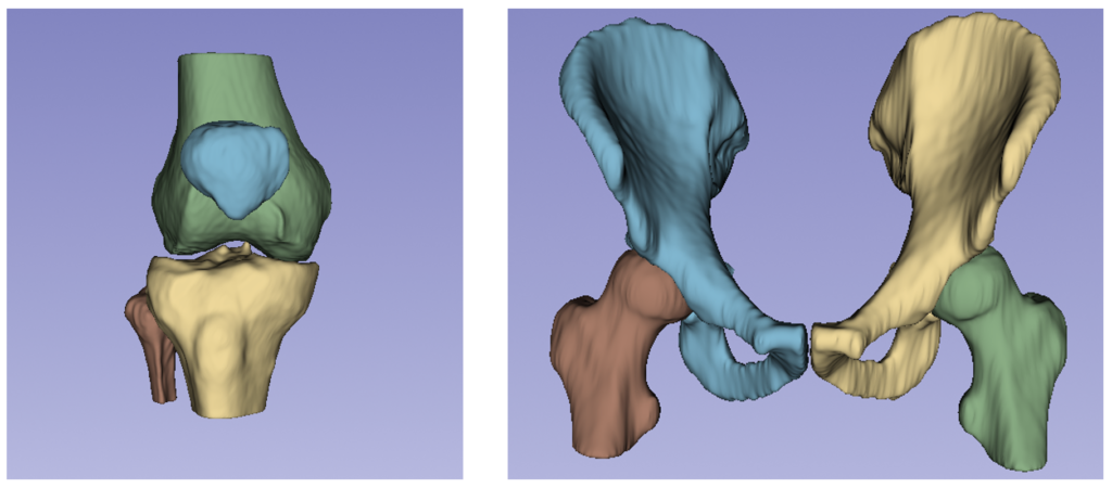
In practical terms, it means identifying the contours, surfaces, and boundaries of bones within a medical image. For example, in an orthopedic planning workflow, segmentation may be used to isolate the femur, tibia, pelvis, humerus, or other anatomical structures depending on the needs of a clinical case.
Once the bone has been segmented, the information can be used to generate a 3D anatomical model. This model can then support measurements, simulations, surgical planning, implant positioning, and, in projects such as ADAPT, the design of customized implants.
This is particularly important because orthopedic procedures are highly dependent on anatomy. Even small differences in bone shape, alignment, deformity, or joint structure can influence surgical strategy and implant design. Recent literature on AI in orthopedics highlights that orthopedic image analysis has benefited from improved segmentation algorithms capable of delineating structures such as bone, cartilage, muscle, and other elements.
How Is Image Segmentation Used in Orthopedics?
In orthopedics, segmentation helps convert medical images into actionable information.
A surgeon, engineer, or planning system may need to understand the exact shape and sizing of a bone before making decisions about alignment, correction, implant size, implant fit, or surgical approach. Manual segmentation can be time-consuming and dependent on user experience. Automated or AI-assisted segmentation can help make this process faster, more consistent, and more scalable.
For example, segmented bone images can be used to:
- create patient-specific 3D anatomical models;
- support pre-operative planning;
- identify bone deformities or anatomical variations;
- simulate implant positioning;
- support the design of patient-specific implants;
- improve communication between clinicians, engineers, and manufacturers.
The use of 3D imaging-based AI models has demonstrated high levels of accuracy and reproducibility, with performance comparable or superior to expert manual segmentation, while also significantly reducing processing time. These advances support the integration of automated segmentation into clinical workflows for orthopedic planning, quantitative analysis, and implant design. Within the ADAPT project, we describe orthopedic image segmentation as a tool that allows surgeons to better explore patient-specific anatomy without spending excessive time on manual image analysis. The same source also explains that segmentation can help distinguish what is bone from what is not, facilitating the orthopedic planning process. In clinical workflows, segmentation may also support post-operative assessment. In hip and knee applications, AI-powered segmentation has been described as relevant for both pre-operative planning and post-operative evaluation, particularly because anatomical differences between patients can influence treatment decisions and outcomes.
How Does Bone Segmentation Work?
Bone segmentation can be performed manually, semi-automatically, or automatically.
In a traditional manual process, a trained professional outlines the bone structure slice by slice or region by region. While this can be accurate, it may also be slow, repetitive, and difficult to scale.
With AI-based segmentation, the process becomes more automated. Machine learning and deep learning models are trained on large sets of annotated medical images. These annotations teach the model to recognize patterns associated with specific anatomical structures. Over time, the system learns to identify bone boundaries and separate them from other regions of the image.
In simple terms, the AI model learns to answer the question: “Which pixels or voxels belong to this bone?”
Once the bone is identified, the segmented region can be transformed into a digital representation of the patient’s anatomy. Depending on the imaging modality and workflow, this may support 2D analysis, 3D reconstruction, anatomical measurements, or implant design.
Why is bone segmentation important for ADAPT?
For ADAPT, bone segmentation is a key step in the personalized implant workflow. The project aims to develop an integrated ecosystem for producing customized metallic orthopedic implants, connecting medical imaging, segmentation, 3D reconstruction, implant design, additive manufacturing, testing, and quality control.
In this context, segmentation acts as the bridge between the patient’s anatomy and the implant. By identifying the relevant bone structures from medical images, it enables the creation of accurate digital anatomical models. These models can then support the design of implants adapted to each patient’s bone geometry, instead of relying only on standard implant sizes.
Better segmentation can lead to better anatomical models; better anatomical models can support better implant design; and better implant design can contribute to improved fit, biomechanical performance, and patient-specific care.
From Medical Image to Customized Implant
The journey from a medical image to a customized implant involves several connected steps.
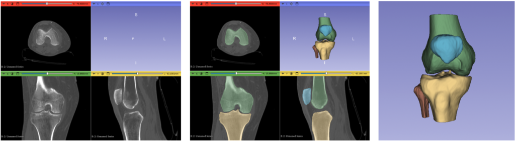
First, the patient undergoes medical imaging, such as CT or MRI. Then, bone segmentation is used to identify and separate the relevant anatomical structures. These segmented structures are converted into 3D models, which can support measurements, simulation, and implant design.
In ADAPT, this digital workflow is directly connected to additive manufacturing, particularly Powder Bed Fusion – Laser Beam technologies. This makes it possible to produce complex, patient-specific implant geometries that would be difficult to achieve through traditional manufacturing methods.
By turning medical images into accurate anatomical models, bone segmentation helps connect clinical data with engineering design. This makes it a critical step in ADAPT’s mission to develop implants that are not only manufactured for the patient, but designed around the patient.