
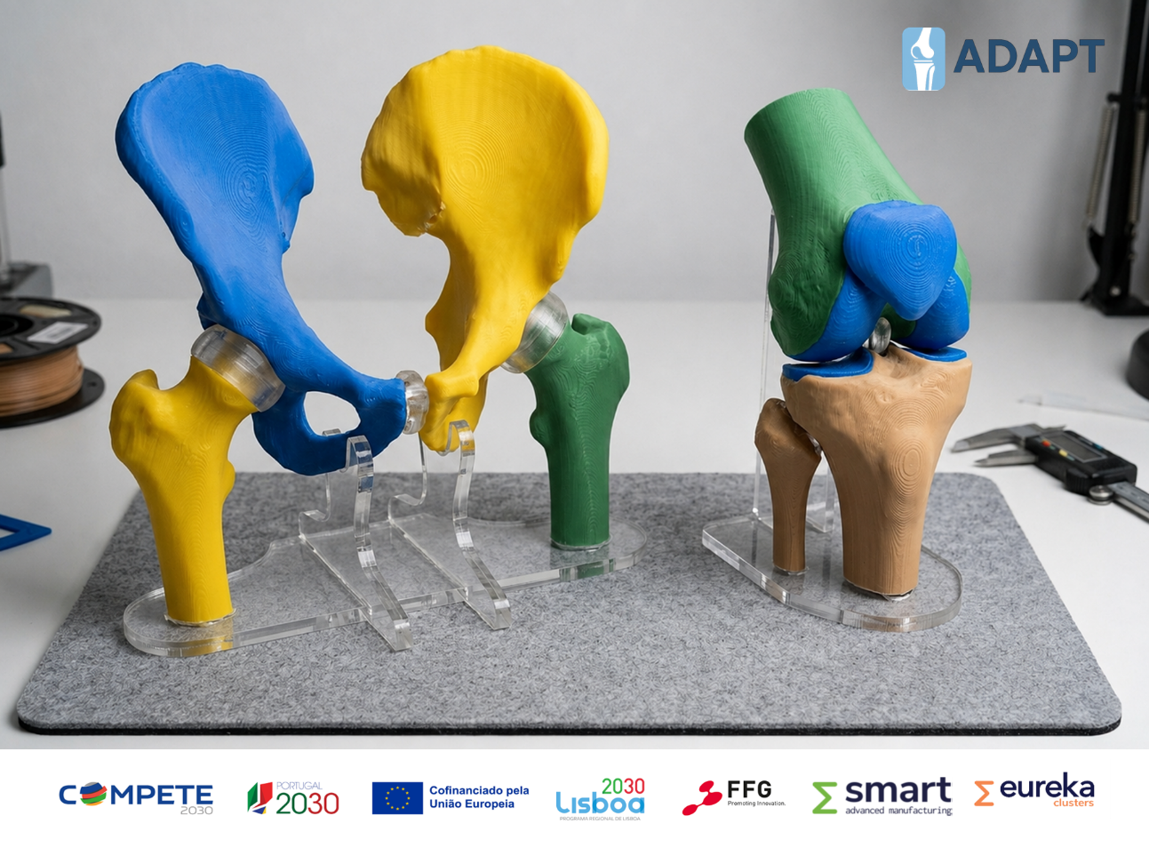
Personalised arthroplasty begins long before the operating theatre. It begins with understanding the patient’s anatomy in detail: the shape of the bone, the degree of deformity, the available bone stock, the alignment of the limb, and the specific challenges that may influence implant positioning and surgical execution. In ADAPT, anatomical bone models are not an isolated digital step: they are part of a broader clinical and manufacturing pathway designed to support more precise, patient-centred and digitally integrated orthopaedic solutions.
From medical imaging to anatomical understanding
In hip and knee arthroplasty, medical imaging is often the first step towards understanding the structural problem. Computed tomography or magnetic resonance imaging allow the clinical and technical teams to collect detailed information on bone morphology, morphometry and topography. However, the clinical value of imaging does not depend only on image acquisition. It depends on how those images are interpreted, segmented and transformed into useful anatomical information.
Through segmentation, two-dimensional imaging data can be converted into three-dimensional digital models of the patient’s bone anatomy. These models allow surgeons, engineers and digital planning teams to move from a flat image to a spatial representation of the joint. For the surgeon, this can support a more intuitive understanding of the case. For the engineer, it provides the anatomical basis for implant design, prototyping and manufacturing decisions.
This is especially relevant in personalised arthroplasty, where the objective is not simply to select the closest standard implant size, but to understand how the implant should respond to the patient’s individual anatomy. The digital bone model becomes a common reference point: a clinically meaningful representation of the patient, and a technical foundation for the steps that follow.
See previous blog post “What Is Bone Segmentation and How Is It Used in Orthopedics?” for more details on bone segmentation: https://www.adapt-smart.eu/what-is-bone-segmentation-and-how-is-it-used-in-orthopedics/
Why bone models matter in hip and knee arthroplasty?
Hip and knee arthroplasty are highly successful procedures, but they remain technically demanding. Even in primary cases, patients can present with substantial anatomical variability. In more complex situations, surgeons may need to manage deformity, bone loss, altered joint geometry, previous trauma or surgeries, unusual morphology or revision-like anatomical challenges. This becomes even more relevant in arthroplasty revision procedures, where previous implants, compromised bone stock and altered anatomy can make preoperative planning essential to reduce uncertainty and support safer surgical decision-making [1,2].
Bone models can be particularly valuable in these contexts. They allow the surgical team to assess the anatomy in three dimensions, identify potential difficulties before surgery and anticipate how the implant may interact with the remaining bone [2]. In the knee, this may include the relationship between the femur, tibia and patella, as well as alignment, bone coverage and joint line considerations. In the hip, it may involve the femoral canal, acetabular anatomy, ante/retroversion, acetabular and femoral offset, bone defects or the restoration of biomechanical parameters.
The clinical importance lies in preparation. A clearer understanding of the patient’s anatomy can support better decision-making regarding implant positioning, sizing and shaping, bone resections, fixation strategy and the sequence of surgical steps. In personalised arthroplasty, this preparation is essential because the surgical plan and the implant concept must be developed together.
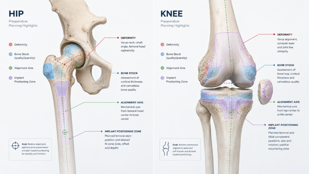
Figure 1 – Three-dimensional bone models can support the assessment of deformity, bone defects, alignment and implant positioning in complex hip and knee arthroplasty planning.
From visualisation to surgical strategy
A bone model is more than a visual object. When properly integrated into surgical planning, it becomes a decision-support tool [3,4].
Digital models allow clinicians and technical teams to visualise the joint from multiple perspectives, measure anatomical parameters and discuss possible surgical strategies. Physical models, produced through 3D printing, can add a tactile and spatial dimension that is particularly useful in complex cases. Holding a realistic model of the patient’s anatomy can help the surgical team appreciate deformity, bone defects or anatomical constraints in a way that is less immediate or understandable on screen [3,5].
In the ADAPT pathway, anatomical models’ prototypes are expected to support visual understanding of the real implant dimensions to be designed and to help identify potential anatomical issues before manufacturing. This does not replace clinical judgement. Rather, it strengthens it by providing a more complete representation of the case.
For surgeons, this may help to respond to questions such as:
- Can the planned implant geometry achieve appropriate bone coverage?
- Are there areas where overhang, under-coverage or conflict with surrounding anatomy may occur?
- Is the available bone stock sufficient for the intended fixation concept?
- Does the anatomy suggest specific risks during bone preparation or implant positioning?
- Would the planned surgical strategy be reproducible in the operating theatre?
These are clinical questions, but they require anatomical precision and technical translation.
Supporting alignment, implant positioning and risk reduction
Alignment remains one of the central themes in hip and knee arthroplasty. It influences lower limb load distribution, joint kinematics, implant stability and long-term performance [1]. While alignment philosophy may vary according to indication, surgeon preference and implant concept, all approaches require a precise understanding of the patient’s anatomy.
The value is not only technical. It is also related to patient safety. By anticipating anatomical constraints before surgery, the clinical team can reduce uncertainty, prepare for specific intraoperative challenges and communicate more clearly with all stakeholders involved in the case. In complex arthroplasty, risk reduction often begins with better planning.
ADAPT does not assume that bone models alone will solve every surgical challenge. However, they can help transform a complex anatomical situation into a structured planning process. This is particularly important when personalised implants are being considered, because every design decision must remain clinically meaningful, surgically feasible and compatible with the patient’s anatomy.
A shared language between clinicians and engineers
Personalised implants require close collaboration between clinical teams, engineers, manufacturing specialists and quality-control experts. Each discipline sees the case through a different lens. The surgeon thinks about exposure, bone preparation, fixation, alignment, soft tissues and intraoperative decision-making. The engineer thinks about geometry, manufacturability, material behaviour, mechanical performance and quality requirements. The manufacturer thinks about process control, tolerances, post-processing and inspection.
Bone models help create a shared language between these perspectives. The pathway cannot be driven by technology alone. The anatomical model must preserve the clinical context and ensure that design and manufacturing decisions remain aligned with surgical objectives.
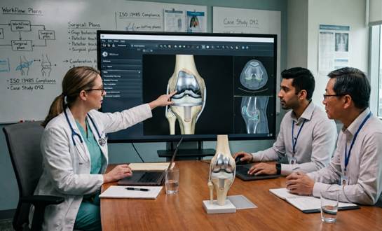
Figure 2 – Anatomical models provide a shared reference for surgeons, engineers and manufacturing teams, helping translate clinical requirements into technically feasible solutions.
Fitting bone models into the wider ADAPT digital chain
Within ADAPT, bone models are part of a larger digital and manufacturing chain. The project addresses the transition from patient-specific data to implant design, additive manufacturing, post-processing, quality control and validation. Anatomical modelling sits at the beginning of this chain, where clinical information is translated into digital geometry.
Bone modelling is one of the foundations of personalised arthroplasty. It supports implant design, but it also supports planning, discussion, prototyping and validation. In the ADAPT project, this is aligned with the broader ambition of developing digital tools, anatomical models and personalised implant pathways that can be more reliable, efficient and clinically relevant.
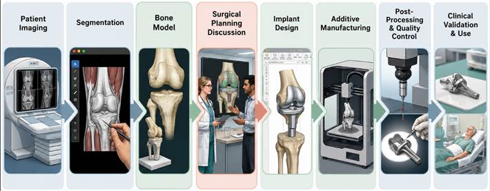
Figure 3 – Bone models sit at the beginning of the personalised implant pathway, linking patient-specific anatomy with design, manufacturing, quality control and clinical validation.
Towards more predictable personalised arthroplasty
Bone models and surgical planning represent a bridge between diagnosis and intervention, between clinical reasoning and digital manufacturing, and between the patient’s anatomy and the final implant concept. They help transform individual anatomical complexity into a structured, multidisciplinary planning process.
Bone models are not merely representations of bone. They are instruments of clinical preparation, communication and safer decision-making. They support the ADAPT ambition to contribute to more precise, patient-centred and digitally integrated orthopaedic solutions for the hip and knee.
References
[1] Rosso, F., Rossi, R., Cottino, U., Dettoni, F., Bruzzone, M., & Bonasia, D. E. (2022). Three-Dimensional Printed Models in Pre-Operative Planning of Complex Primary and Revision Total Knee Arthroplasty. Applied Sciences, 12(19), 9618.
[2] Maryada, V. R., Mulpur, P., Eachempati, K. K., Annapareddy, A., Prasad, V. B. N., & Reddy, A. G. (2022). Pre-operative planning and templating with 3-D printed models for complex primary and revision total hip arthroplasty. Journal of Orthopaedics, 34, 240-245.
[3] Jiang, M., Chen, G., Coles‐Black, J., Chuen, J., & Hardidge, A. (2020). Three‐dimensional printing in orthopaedic preoperative planning improves intraoperative metrics: a systematic review. ANZ journal of surgery, 90(3), 243-250.
[4] Alemayehu, D. G., Zhang, Z., Tahir, E., Gateau, D., Zhang, D. F., & Ma, X. (2021). Preoperative planning using 3D printing technology in orthopedic surgery. BioMed Research International, 2021(1), 7940242.
[5] Morgan, C., Khatri, C., Hanna, S. A., Ashrafian, H., & Sarraf, K. M. (2020). Use of three-dimensional printing in preoperative planning in orthopaedic trauma surgery: A systematic review and meta-analysis. World journal of orthopedics, 11(1), 57.
